Dr. Yan Sze Wan and Dr Yew Wing Wai
TB and Chest Unit, Grantham Hospital
Case History and Physical Examination
A young man aged thirty-seven presented himself fifteen years ago with cough since his childhood. He reported frequent bronchitic symptoms with expectoration of whitish sputum. In one of the episodes, he noticed some blood tinge.
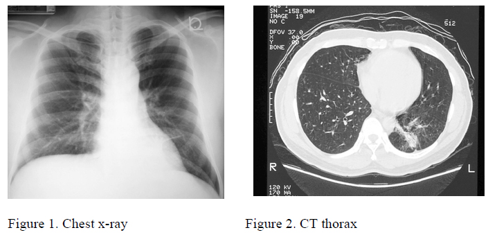
On physical examination he had normal body built. There was no clubbing of fingers. Chest examination was unremarkable alongside with other systems. Chest x-ray showed a retrocardiac shadow (Figure 1).
Investigations
Blood tests showed normal haemoglobin. There was no systemic leucocytosis and the differential count showed normal level of eosinophil. Biochemistry for liver and renal function tests was unremarkable. Multiple sputum specimens showed negative findings for cytology examination and no acid fast bacilli were identified. CT thorax revealed a shadow at posterior basal segment of left lower lobe (figure 2).
Further investigation with fibreoptic bronchoscopy showed no endobronchial lesion. Transbronchial biopsy at left lower lobe was unrevealing.
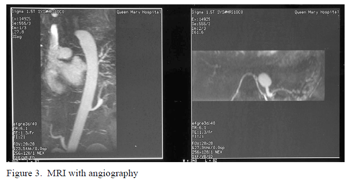
More advanced imaging study with 3–dimensional Magnetic Resonance Angiography (MRA) demonstrated the arterial supply from the descending thoracic aorta to the left lower lobe (figure 3)
Diagnosis: Sequestration of Lung
Literature Review
Please refer to Question 3 of “Practical Corner” section on page 26